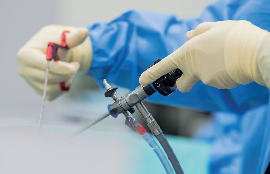
Exploring technological advances in patient treatment


Mays Cancer Center is at the forefront of technology-assisted surgical treatments that are improving patient recovery.
Robotic kidney cancer surgery shows desirable outcomes
Kidney cancer is not always confined to the kidney. In advanced cases, this cancer invades the body’s biggest vein, the inferior vena cava (IVC), which carries blood out of the kidneys back to the heart. Via the IVC, cancer may infiltrate the liver and heart.
In a study published in the Journal of Urology, researchers from the Mays Cancer Center and the Department of Urology at UT Health San Antonio show that robotic IVC thrombectomy (removal of cancer from the inferior vena cava) is a highly safe and effective alternative approach to standard open IVC thrombectomy. During surgery, the affected kidney is removed along with the tumor.
The open surgery requires an incision that begins two inches below the ribcage and extends downward on both sides of the ribcage.
“It looks like an inverted V,” said Dharam Kaushik, MD, urologic oncologist and senior author of the study. Next, organs that surround the IVC, such as the liver, are mobilized, and the IVC is clamped above and below the cancer. In this way, surgeons gain control of the inferior vena cava for cancer resection.
“Open surgery has an excellent success rate, and most cases are performed in this manner,” Kaushik said. “But now, with the robotic approach, we can achieve similar results with smaller incisions.”
The study is a systematic review and meta-analysis of data from 28 studies that enrolled 1,375 patients at different medical centers. Of these patients, 439 had robotic IVC thrombectomy and 936 had open surgery. Kaushik and his team collaborated with Memorial Sloan Kettering Cancer Center, New York; Cedars-Sinai Medical Center, Los Angeles; and the University of Washington, Seattle, to perform this study.
“We pulled the data together to draw conclusions because, before this, only small studies from single institutions had been conducted to compare the IVC thrombectomy approaches,” Kaushik said. “In more than 1,300 patients, we found that overall complications were lower with the robotic approach and the blood transfusion rate was also lower with this approach.”
Among the outcomes:
- Fewer blood transfusions: 18% of robotic patients required transfusions compared to 64% of open patients.
- Fewer complications: 14.5% of robotic patients experienced complications such as bleeding compared to 36.7% of open thrombectomy patients.
“That tells us there is more room for us to grow and refine this robotic procedure and to offer it to patients who are optimal candidates for it,” Kaushik said.
MRI technique improves detection of prostate cancer
An MRI scan called restriction spectrum imaging greatly improves the detection of prostate cancer progression, according to a study published in the Journal of Urology by researchers at the Mays Cancer Center.
The technique, called RSI-MRI, improves the pictures produced from more traditional scanning to reveal biological features, or biomarkers, showing changing conditions of existing prostate cancer patients. By detecting changes in patients more accurately, physicians can move ahead more quickly with potentially life-saving treatment.
In a study enrolling 123 patients from January 2016 to June 2019, researchers developed a targeted, short-duration RSI-MRI scan of less than five minutes that can be added to standard multi-parametric magnetic resonance imaging, or mpMRI, which has been more effective in diagnosing prostate cancer by detecting changes in existing patients.
The study participants on “active surveillance,” or observation, underwent both mpMRI and RSI-MRI scanning reviewed by a urological radiologist for lesions, followed by an MRI-guided prostate biopsy by a urologist.
The RSI-MRI analysis generated biomarkers called restricted signal map (RSM) values, which were then compared with more traditional measures, for detection of worsening grades of prostate cancer. That analysis revealed better accuracy, including a higher level of true-positive lesions and fewer false-positives.
“We found that the RSI-MRI scan provides a more sophisticated analysis and a more sensitive and specific imaging biomarker of aggressive prostate cancer,” said Michael A. Liss, MD, MAS, FACS, a urologic oncologist at the Mays Cancer Center, and the study’s senior author.
“The findings show that using a simple acquisition technique of measuring and storing data can substantially enhance MRI,” Liss said. “Also important is that RSI-MRI acquisition does not need hardware or contrast material and can be deployed within the current MRI workflow.”
Enhanced robotics system speeds recovery for testicular cancer patients
Testicular cancer mainly affects young men, and its treatment requires multidisciplinary care and significant coordination between surgeons and medical oncologists. The disease itself has excellent cure rates. However, it commonly spreads to abdominal lymph nodes.
With an innovative approach using an enhanced robotics system, surgeons at the Mays Cancer Center can now treat testicular cancer patients without the traditional open surgery. The process includes inserting cameras and surgical instruments inside the abdomen and controlling them using robotic systems to remove cancerous lymph nodes.
“This is great news for testicular cancer patients,” said Ahmed Mansour, MD, MRCS, urologic oncologist.
Not that long ago, standard surgery required 10-inch incisions across the abdomen and led to lengthier and more painful hospital stays and recovery. The newer procedure is called robotic retroperitoneal lymph node dissection, or robotic RPLND.
“It is a challenging procedure,” Mansour said. “After chemotherapy, the lymph nodes become closely attached to the major blood vessels, and taking them out involves risk of injury.”
For that reason, robotic RPLND largely has been reserved for cases in which chemotherapy hasn’t been done or nodes aren’t visibly enlarged. However, Mays Cancer Center researchers hit upon a novel approach, developing technical improvements to make it safer, more effective and applicable in new indications.
“With some technical modifications, we are able to mimic open surgery in patients with bulky lymph nodes who have already received chemotherapy, Mansour said. Precisely maneuvering instruments with the help of robotics requires only small, half-inch incisions. Moreover, robotic RPLND now can be performed to remove nodes from both sides of the body in one setting.
While both RPLND procedures remain challenging and continue to take eight to 10 hours to perform, patients now can leave the hospital after two days, and the smaller incisions heal faster and with less scarring. Evaluation of long-term outcomes of patients undergoing robotic RPLND is underway.
Read more stories from our 2022 Annual Report.
Explore our Cancer Surgery and Cancer Research and Clinical Trials Programs.

 Close
Close